AI microscopy for everyone

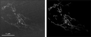
The restoration DL model receives as input a low signal to noise ratio (SNR) image (left) and output a high SNR image (right). Image depicts a cell with fluorescently labelled mitochondria imaged by iSIM (super resolution microscopy).
DRVISION announces the launch of Aivia Cloud, the pioneering, universally usable artificial intelligence platform for scientific imaging applications.
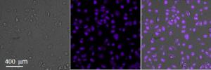
BELLEVUE, WA, USA, November 12, 2018 /EINPresswire.com/ -- DRVISION announces the launch of Aivia Cloud, the pioneering, universally usable artificial intelligence (AI) platform for scientific imaging applications. The unique cloud-desktop hybrid platform for training and deployment of deep learning (DL) models is the result of an 18 month long joint industry-academy R&D project which involved 25 experts in AI, life sciences, microscopy and image analysis.DL, a type of AI technology, is revolutionizing many areas of our daily lives and is now starting to be adopted by the life sciences imaging community. In the past 18 months, a growing volume of DL-based scientific reports have shown that DL-powered tools can help solve microscopy and image analysis problems. DL-powered tools can be used to improve image resolution, to increase image signal to noise ratio, automatically identify objects of interest, and predict the localization of subcellular features in microscopy images (reference 1-4). These approaches allow scientists to run previously impossible imaging experiments and quickly extract meaning from raw data. Aivia Cloud users can directly apply any of the three DRVISION-trained models (see images for examples), train new DL models exclusively with their own data or choose to augment one of the pre-trained models by transfer learning. Aivia Cloud leverages Google Cloud Platform’s state-of-the-art computing infrastructure, as well as TensorFlow, Keras and Python.
"AI has the potential to redefine the limits of microscopy which would greatly accelerate the rate of knowledge creation. However, due to the massive shortage of deep learning experts the wider imaging community has not been able to benefit from this type of tech. In collaboration with our academic partners, our in-house experts have created a pipeline that can be used by any scientist, no coding or AI experience needed. Aivia Cloud is set to democratize the use of AI for a myriad of image-based applications for both life and material sciences" says Luciano Lucas (PhD), Executive VP at DRVISION.
What are the factors limiting the adoption of DL technology for life sciences imaging applications:
DL-based technology is currently only used by small group of pioneering research groups. This is due to four major practical hurdles: (1) the requirement for multiple highly specialized and disjointed software libraries or tools to cover the entire DL train-apply workflow. This step often requires users to reformat or resave their data in several different file formats; (2) the need to have extensive expertise to finetune the training process; (3) the need to access expensive, high-performance hardware to train DL networks, and (4) the difficulty and time-consuming properties of ground truth (GT) creation.
Aivia Cloud removes the first three bottlenecks and accelerates the generation of GT data. The first issue is resolved by creating a fully integrated platform which covers the whole pipeline, from the creation of ground truth annotations to training and applying DL models. The second issue is addressed by developing and testing hyperparameter optimization procedures (patent pending) which are robust for the three application types initially supported. The third issue is solved by integrating and leveraging a world class cloud computing platform – the Google Cloud Platform. The final issue is solved by Aivia’s patented machine learning technology, parameter-free image segmentation as well as Aivia’s non-ML high performance image processing tools.
Aivia Cloud was publicly launched in San Diego, California, USA, at the world’s largest neuroscience conference: the annual meeting of the Society for Neuroscience (SfN) that brings together over 30K attendees. For more information and to request early access to Aivia Cloud, contact Luciano Lucas (lucianol@drvtechnologies.com, +1 425 773 1548).
Presentation introducing Aivia Cloud (and Aivia 8): https://youtu.be/VP9QlRfrdZA
More information:
Aivia Cloud: www.drvtechnologies.com/aivia-cloud
Aivia 8 (also launched): https://www.drvtechnologies.com/aivia8
Aivia (overview of all functionality): https://www.drvtechnologies.com/aivia
Deep Learning in Aivia: https://www.drvtechnologies.com/deep-learning-aivia
About DRVISION Technologies:
DRVISION works with scientists and engineers at the technological frontier, and pioneers image based decision technologies that propel major breakthroughs in the life science, electronics and materials industries. DRVISION is a technological innovator with 50 issued US patents, and commercial interests in X-ray inspection, survey, search / alignment, video inspection and life sciences. DRVISION makes and markets Aivia microscopy image analysis software. Aivia development is partially funded by the National Institutes of Health (NIH) under multiple Small Business Innovative Research (SBIR) programs worth over $10M. For more information, visit www.drvtechnologies.com.
Image credits
Restoration and prediction DL model example: Dr. Hari Shroff, Dr. Jiji Chen, Mr. Yijun Su (NIBIB/NIH, Bethesda, USA)
Electron microscopy segmentation DL model example: Prof. Rachel Wong and Dr. Wan-Qing Yu (University of Washington, Seattle, USA).
References
1. Riverson Y, et al., Optica, Volume 4, Issue 11, (2017)
2. Weigert M, et al., Biorxiv, (2018)
3. Turaga SC, et al., Neural Computation, Volume 22, Issue 2, (2010)
4. Christiansen EM, et al., Cell, Volume 173, Issue 3, (2018)
Luciano Lucas
DRVISION Technologies
+1 425-773-1548
email us here
Legal Disclaimer:
EIN Presswire provides this news content "as is" without warranty of any kind. We do not accept any responsibility or liability for the accuracy, content, images, videos, licenses, completeness, legality, or reliability of the information contained in this article. If you have any complaints or copyright issues related to this article, kindly contact the author above.


