arivis AG Introduces Newest Virtual Reality (VR) Software for Analysis of Microscope 3D and 4D Image Data

The latest version of arivis InViewR provides efficient, accurate, and interactive proof reading, editing, and segmentation of real, 3D images in Virtual Reality (VR). Image sources include light & electron microscopes, CT's & MRI's
InViewR's proof-editing and segmentation features will save me days compared to trying to manually complete my work on a desktop. It's a game changer.”
MUNICH, GERMANY, November 10, 2017 /EINPresswire.com/ -- arivis AG will introduce its latest version of InViewR software at the 2017 Society for Neuroscience Meeting (SfN) November 11th – 15th in Washington, DC. InViewR displays real image data in VR by utilizing patent pending direct volume rendering techniques with no need to convert data or make surface models. The latest InViewR sets new standards for analyzing life science research images by enabling efficient, accurate, and interactive proofreading, proof-editing, and de-novo segmentation of multi-dimensional images from virtually any source instrument.— Elizabeth R.
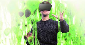
In VR the user can enter their data and position one’s viewpoint within the data itself, thus breaking through limitations of viewing 3D/4D images on a 2D desktop screen, like constantly having to turn the image with a mouse. With natural movements of the head and body a user can move freely to inspect and interact with an image from any angle and position without limitation. Users are instantly able to comprehend and internalize information about important relationships between structures within an image. These new insights, many impossible to perceive on a desktop system, can be used to evaluate existing hypotheses, create new ones, and generate appropriate data analysis strategies.
Freed from being tethered to a mouse like on a desktop computer and with depth perception equivalent to the real word, a person’s hands are unencumbered to simply reach into the data to precisely and intuitively mark, measure, classify, edit, and segment. A cumbersome and frustrating process that on a desktop involves, multiple turns of the dataset, changing tools multiple times, guessing at which object is being selected, and iteratively positioning from different angles is reduced to simply reaching out and pulling a trigger or pushing a button.
“InViewR makes impossible or enormously time consuming segmentation tasks possible and reasonable to undertake” states Michael Wussow, Vice President Sales and Marketing, Imaging. “Users are able to take images 80% segmented with automatic algorithms to 100% completion by using sculpting tools to add to, remove from, delete and join segments. Manual segmentation tasks that on a desktop took researchers weeks or months are completed in hours or days with increased accuracy using semi-automatic, point and shoot, and manual painting tools.”
Because segmented data is overlaid on the original volume data in InViewR, statistical properties of segments can be calculated in real time based on the original data even as segments are modified, added, and deleted. Statistics on position, size, shape, and per color channel intensity values are calculated. Those statistics can be viewed in VR space, in a table in the desktop portion of the application, and can be exported to excel or other statistical programs for analysis. The segments themselves can be seamlessly passed to a desktop program like arivis Vision4D for further analysis or can be exported as object files to be used in other programs.
InViewR can be used to manually segment structures otherwise impossible to segment algorithmically. Structures that cross each other would normally confuse automatic algorithms. However, our brain is adept at figuring out which piece of a crossing structure is a continuous part of the same structure after the crossing point. Because it is possible to visually follow the path of the structure of interest in VR we are able to paint the structure to segment it independently of other parts of the data. The generated segments can be transferred to desktop software like arivis Vision4D which can mask out just that portion of the original data to create a color channel for each segmented region. It is possible to interactively color and turn on and off segmented portions of the original data for presentation and analysis.
InViewR is a part of the arivis imaging platform. As such, image and segment data is seamlessly transferred between arivis applications. No matter the stage in the visualization and analysis process a customer is at, they have the flexibility to choose the software tool appropriate for their needs. Customers can take advantage of the unique strengths of each tool in the platform.
With over 10 years of development and regarded as the standard for working with large images, the desktop solution arivis Vision4D may be the preferred tool for automatic, algorithmic segmentation. arivis InViewR may be the best tool for proofreading and proof-editing. arivis WebView, a client / server application that is accessible via any standard web browser, may be the ideal tool for harnessing the computer power of servers for batch processing images and then sharing the results with remote clients and collaborators. No matter the path a customer chooses the arivis platform allows customers to get the results they need seamlessly.
Demonstrations of this software will be available at arivis Booth 3015 and Carl Zeiss Booth 2923 during the SFN exhibition. More information, including movies, can be found at www.arivis.com/vr, emailing info@arivis.com or calling 1-800-377-6962.
About arivis AG
arivis specializes in big image data and compliance software for the life, health- and material sciences. Its software enables users to visualize, analyze, distribute and manage multi-terabyte sized files and multi-dimensional (2D, 3D, 4D, 5D) image datasets that are created by microscopes or scanners. arivis software solutions also help customers to meet regulatory, quality and compliance requirements in research, clinical trials, approval, and maintenance of medical devices and medicinal products. arivis serves the global life science communities from their headquarters in Munich, Germany, with subsidiaries in the United States. More info at www.arivis.com
Michael Wussow
Arivis AG
651-336-4600
email us here
See arivis InViewR in action. Watch a demonstration of Visualization, Segment Proofreading & Proof-editing, and Selective Visualization Tools


